Moving beyond traditional 2D assays, Reprotox Biotech provides a physiologically relevant 3D mini-testis model that recapitulates the complex testicular niche. Our multi-cellular co-culture system includes Spermatogonia, Sertoli, and Leydig cells embedded in a specialized extracellular matrix (ECM) to mimic in vivo morphology and paracrine signaling. These animal-free reproductive toxicity services are aligned with New Approach Methodologies (NAMs) and support mechanism-informed screening in male reproductive toxicology.
ReproTox has developed an animal-free 3D testicular cell co-culture system (3D-T®Repro) from a combination of testicular cell lines. This model has gone through extensive biomarker and morphology testing to ensure that it reflects in vitro conditions and provides affordable, humane, and efficient methods for reproductive and developmental toxicity screening.
ReproTox has developed a proprietary technology to establish in vitro 3D testicular models at different stages of spermatogenesis from mice, rats, and canine. We derive testicular cells from neonate, juvenile, and adolescent sources to establish testicular-like organoids representing differential stages of spermatogenesis.
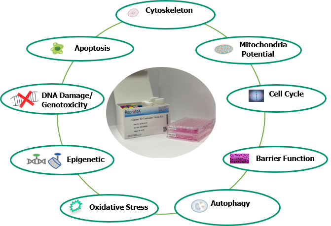
This organoid culture recapitulates many in vivo features and enables assessment of differentiation and mitosis. An extensive panel of toxicological endpoints is assessed in multiple cell types and at multiple time points using High-Content Screening (HCS). By combining these endpoints with exposure levels, the system delivers a sensitive and specific prediction of repro-toxicity.
3D-T®Repro_EX-Vivo provides an innovative technology to culture seminiferous tubules and enable spermatogenesis in vitro. The technology preserves the original structure of the seminiferous tubules, and Sertoli cells remain polarized—crucial for germ cell differentiation—allowing the study of both mitotic and meiotic phases.
ReproTox is at the forefront of cell-based High-Throughput (HT) and pathway-based High-Content Analysis (HCA) for reproductive toxicity evaluations. We provide fully automated HT screening and high-content analysis, plus access to our reproductive toxicity database (150 compounds). Fluorescence-based microplate assays enable rapid, quantitative readouts suitable for High-Content Analysis.
| Assay No | Name/Species | Endpoint | Cell Type | Price |
|---|---|---|---|---|
| CS-SCs | Canine Sertoli Cell | Viability/Cytotoxicity, Multiplexed High-Content analysis | Canine, Primary Sertoli Cell | Quote |
| MU-SCs | Mouse Sertoli Cell | Viability/Cytotoxicity, Multiplexed High-Content analysis | Mouse, Sertoli Cell Line | Quote |
| MU-LCs | Mouse Leydig Cell | Viability/Cytotoxicity, Multiplexed High-Content analysis | Mouse, Leydig Cell Line | Quote |
| MU-SPs | Mouse Spermatogonial Cell | Viability/Cytotoxicity, Multiplexed High-Content analysis | Mouse, Spermatogonial Cell | Quote |
| HU-SCs | Human Sertoli Cell | Viability/Cytotoxicity, Multiplexed High-Content analysis | Human Primary Sertoli Cells | Quote |
| HU-LCs | Human Leydig Cell | Viability/Cytotoxicity, Multiplexed High-Content analysis | Human Primary Leydig Cells | Quote |
| 3D-T®Repro_Primary Canine | Canine Organoid | Viability/Cytotoxicity, Multiplexed High-Content analysis | Canine Primary Testicular Cells | Quote |
| 3D-T®Repro_Primary Mouse | Mouse reproductive 3D model | Viability/Cytotoxicity, Multiplexed High-Content analysis | Mouse primary testicular cells | Quote |
| 3D-T®Repro_Primary Rat | Rat reproductive primary cell 3D model | Multiplexed High-Content analysis | Rat primary testicular cells | Quote |